- +49 201 43 70 97 0
- info@pakumed.de
- Mon - Fri: 8:30 - 17:00
Yes ANOKRYO® can be used during these periods without any problems. As opposed to other gel and ointment preparations, there are no side effects.
Depending on symptoms approx. 3-4 x daily.
This is not a problem. The important thing is that the ANOKRYO® rod be stored in the freezer. Normal refrigerator temperatures are insufficient.
No! Due to its water solubility it would freeze. The gel is applied prior to use.
No there are no adverse effects (see IFU).
Yes without any problems. Anokryo is also helpful in treating constipation
ANOKRYO® is available in the pharmacy or may be ordered there on a non-prescription basis. You may order it at this internet site.
You can purchase the product at this internet website. Modes of payment are explained at the site.
ANOKRYO® -combi-set: PZN 03941536.
ANOKRYO® -gel: PZN 07470157.
Since 1990
Yes the instructions for usage have been translated in various languages.
No. There is only one production size.
The rod is made of tissue compatible plastic.
Puncture of the port system requires the use of a special needle with a bevelled tip. These needles are the only way to avoid punching defects on the silicone membrane. The needles are available in different lengths and bore diameters depending on which substances are to be injected.
The port needle is used to puncture the port chamber. The needle tip pierces a silicone membrane of the port.
Only port needles have a special needle tip. Only with these needles can punching defects on the silicone membrane be avoided.
It is doubly safe as it has a special safety system designed to prevent needlestick injuries.
The operator will find all details about the safety system in the respective instruction manual.
Yes, but we recommend our 100% guaranteed punch-free needles.
SFN stands for “punch-free needle”.
A “Huber” cannula (named after the inventor) is a commonly used port needle for accessing the port.
The tip is different from PakuMed’s SFN port needle.
The choice of size depends, among other things, on the patient’s anatomy, the location and type of application and is the decision of the doctor.
The port cannula is for single use and can remain in the port for up to 48 hours (recommended). Thus, this can be used to administer medicines and fluids, such as artificial food, over a longer period of time.
Example of a procedure:
The patient can sit or lie down comfortably.
The piercing is done under germ-free conditions (sterile) so that the port system retains its function for a long time and does not become infected.
The patient should not speak or breathe in the direction of the port catheter system during lancing.
Important: A cannula is intended for single use and should not be left lying around for longer than 48 hours (risk of infection otherwise increased).
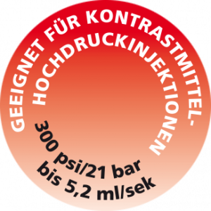
This port catheter system in combination with suitable cannulae enables a high-pressure infusion of contrast media into the central circulatory system. The system is suitable to achieve a flow rate of 5ml/s at a maximum pressure of 21 bar (300 PSI). The pressure stability is tested accordingly and the indication of the suitable port systems or cannulas can be found with us, e.g. by a separate marking for the suitable articles.
A port catheter system, also called a port, port catheter or port system for short, is a long-term access option to the vascular system. It is implanted under the protection of the skin. The implantation is done through a relatively small operation, which can also be done on an outpatient basis under local anaesthesia. The port catheter system consists of the titanium port chamber and a catheter. The titanium port chamber is sealed with a silicone membrane. For the administration of medication, so-called artificial nutrition or blood sampling, a special port cannula is inserted through the skin and through the silicone membrane into the titanium port chamber.
The port catheter system provides permanent access to the vascular system, whereby the catheter is placed in a peripheral blood vessel, usually a vein. This allows access to the vascular system at any time and avoids straining the peripheral veins through multiple punctures. Protecting the skin reduces the risk of infection.
In most cases, it is used for tumour patients who need repeated chemotherapy. The quality of life is significantly improved because the patient’s freedom of movement is not restricted and piercing is much easier and less risky. Normal activities are possible, even sports, swimming, cycling, etc. The implanted port chamber is only slightly raised under the skin and is usually visually unnoticeable. However, it can be easily felt.
The tip of the port catheter is usually in a large vein in front of the heart entrance, the superior vena cava.
The catheter is usually inserted through the subclavian or internal jugular vein or the external jugular vein.
The port chamber is usually located above the right costal arch.
However, other localisations are also possible. This is decided by the doctor.
The implantation of the port catheter system takes place in the clinic as an inpatient or also as an outpatient within about one hour.
Up to two skin incisions are made and the port is pushed under the skin and the catheter is inserted into the large vein.
The patient can choose between a general or local anaesthetic.
The decision should be discussed with the doctor in charge.
During the first few days after the operation you may experience a slight pain or swelling of the skin area, this is normal.
If this persists, reddening of the skin, fever or bleeding occurs, the patient should consult a doctor to avoid complications.
The skin incisions are sutured or covered with sterile plaster strips. These can be removed painlessly from the 8th day after the operation.
When a bandage is no longer necessary, the patient can shower and later bathe after complete healing.
Within the first 14 days, the patient should still take it easy.
The port can be used immediately.
All the drugs that are approved for intravenous therapy can be given via the port catheter system.
This can be:
Infusion solutions such as NaCl, glucose, electrolytes, cytostatics, antibiotics, painkillers such as morphine, parenteral nutrition such as fats, amino acids (protein) and blood products such as platelet concentrates.
The corresponding instructions for use and care recommendations must be observed.
The patient should also carry a patient passport with him/her at all times, if possible, in which all important information is documented.
The doctor will decide when to give which medicine.
It is very important to rinse the port catheter system thoroughly after each administration of medication, otherwise it could become blocked.
Important: The port chamber is not a reservoir and is not filled with blood or medication! After rinsing, it may only contain the appropriate rinsing solution (e.g. saline/heparin solution).
Example of a procedure:
The patient can sit or lie down comfortably.
The piercing is done under germ-free conditions (sterile) so that the port system retains its function for a long time and does not become infected.
The patient should not speak or breathe in the direction of the port catheter system during lancing.
Important: A cannula is intended for single use and should not be left lying around for longer than 48 hours (risk of infection otherwise increased).
The patient should generally observe him/herself daily and pay particular attention to the area around the port catheter system, as well as any swelling of the lymph nodes under the armpits; changes should be reported to the doctor and nurse.
When not in use, flush approx. every 3 months to prevent occlusion of the port catheter system. This is usually done by the family doctor as part of the regular blood checks.
If the port needle is in place, additional daily checks are carried out by the doctor and nursing staff, who check that the port needles are correctly positioned and fixed in place.
Regular flushing is also necessary when giving infusions.
This is particularly necessary before and after the administration of blood samples and after different infusions (interactions) given one after the other.
The doctor and/or nurse flushes the catheter with a syringe filled with at least 10 ml NaCL 0.9 % (10-30 ml saline solution).
Only syringes larger than 10 ml are recommended for each bolus administration.
The same procedure is required when pulling the needle.
This protects the catheter system in the long term.
| Problem | Possible cause | What can be done about it? |
| Higher resistance when injecting, no aspiration of blood. | The catheter tip may rest against the vessel wall. |
|
| Port and catheter cannot be flushed under normal pressure. No blood can be drawn. | Port catheter closure | In all these cases, contact the family doctor immediately. The doctor will then discuss the necessary steps together with the patient. |
| Physical discomfort, pain and possibly swelling in the area of the shoulder, neck and arm | Vein thrombosis | |
| Pain and/or redness at the implantation site, fever or an unusual discharge at the injection site | Infection of the port pocket | |
| Burning pain after infusion of a drug, possibly in combination with blistering of the skin or swelling in the area of the port pocket | Leaky system, the drug seeps into the surrounding tissue. |
| Problem | Possible cause | What can be done about it? |
| Rejection of the port by the body | Incompatibility of the materials | In all these cases, contact the family doctor immediately. The doctor will then discuss the necessary steps together with the patient. |
| Spontaneous slippage of the port | Inappropriate fixation of the port during implantation | |
| Twisting or slipping of the port as a result of unusual movements | Inappropriate fixation of the port during implantation | |
| Pinching of the catheter between the clavicle and the first rib | Individual, anatomical position. td, Surgical technique. |

This port catheter system in combination with suitable cannulae enables a high-pressure infusion of contrast media into the central circulatory system. The system is suitable to achieve a flow rate of 5ml/s at a maximum pressure of 21 bar (300 PSI). The pressure stability is tested accordingly and the indication of the suitable port systems or cannulas can be found with us, e.g. by a separate marking for the suitable articles.
The TITAN-PORT APH (extracorporeal apheresis) is a fully implantable port catheter system as an access option for performing extracorporeal apheresis
This allows access to the vascular system at any time and avoids straining the peripheral veins through multiple punctures. Protecting the skin reduces the risk of infection.
The quality of life is significantly improved because the patient’s freedom of movement is not restricted and piercing is much easier and less risky. Normal activities are possible, even sports, swimming, cycling, etc. The implanted port chamber is only slightly raised under the skin and is usually visually unnoticeable. However, it can be easily felt.
The port catheter is usually located in a large vein in front of the entrance to the heart, the superior vena cava.
The catheter is usually inserted via the internal or external jugular vein.
The port catheter tip is located 2-4 cm in the right atrium and the port chamber is usually above the right costal arch.
However, other localisations are also possible. This is decided by the doctor.
The port catheter system enables optimum flow thanks to its streamlined design with the use of straight port cannulas.
Due to the optimized design, flows of up to 250 ml/min can be achieved during apheresis.
The port catheter system is implanted in hospital or on an outpatient basis within around one hour.
Up to two skin incisions are made, the port is inserted under the skin and the catheter is inserted into the large vein.
The patient can choose between general or local anesthesia.
The decision should be discussed with the responsible doctor.
A slight pain or swelling of the skin area may occur in the first few days after the operation; this is normal.
If this persists for a longer period of time, reddening of the skin, fever or bleeding occurs, the patient should consult a doctor to avoid complications.
The skin incisions are sutured or covered with sterile plaster strips. These can be removed painlessly from the 8th day after the operation.
If a dressing is no longer necessary, the patient can shower and later, after complete healing, bathe.
The patient should take it easy physically for the first 14 days.
The port can be used for therapy after approx. 7 days once the wound has healed.
Recommendation: as a blocking agent e.g. citrate, taurolidine citrate, heparin, urokinase/streptokinase
(diluted heparin is not recommended due to its short half-life)
These are recommendations; they must always be adapted to the patient’s general coagulation situation and are the responsibility of the attending physician.
(carry out an X-ray check beforehand if necessary)
It is mandatory to use only punch-free port cannulas
Example of a procedure:
The patient can sit or lie down comfortably.
The port is inserted under aseptic conditions (sterile) so that the port system retains its function for a long time and does not become infected.
The patient should not speak or breathe in the direction of the port catheter system during insertion.
Important: A cannula is intended for single use and should not be left lying around for longer than 48 hours (risk of infection otherwise increased).
The patient should generally observe themselves daily and pay particular attention to the area around the port catheter system and any swelling of the lymph nodes under the armpits; any changes should be reported to the doctor and nurse.
If not in use, flush approx. every 3 months to prevent the port catheter system from becoming blocked. This is usually done by the family doctor as part of the regular blood checks.
If the port needle is in place, additional daily checks are carried out by the doctor and nursing staff, who check that the port needles are correctly positioned and fixed in place.
Regular rinsing is also necessary when administering infusions.
This is particularly necessary before and after the administration of blood samples and after different infusions (interactions), which are given one after the other.
The doctor and/or nurse flushes the catheter with a syringe filled with at least 10 ml NaCL 0.9 % (10-30 ml saline solution).
Only syringes larger than 10 ml are recommended for each bolus administration.
The same procedure is also required when withdrawing the needle.
This protects the catheter system in the long term.
| Problem | Possible cause | What can be done about it? |
| Higher resistance when injecting, no aspiration of blood. | The catheter tip may rest against the vessel wall. |
|
| Port and catheter cannot be flushed under normal pressure. No blood can be drawn. | Port catheter closure | In all these cases, contact the family doctor immediately. The doctor will then discuss the necessary steps together with the patient. |
| Physical discomfort, pain and possibly swelling in the area of the shoulder, neck and arm | Vein thrombosis | |
| Pain and/or redness at the implantation site, fever or an unusual discharge at the injection site | Infection of the port pocket | |
| Burning pain after infusion of a drug, possibly in combination with blistering of the skin or swelling in the area of the port pocket | Leaky system, the drug seeps into the surrounding tissue. |
| Problem | Possible cause | What can be done about it? |
| Rejection of the port by the body | Incompatibility of the materials | In all these cases, contact the family doctor immediately. The doctor will then discuss the necessary steps together with the patient. |
| Spontaneous slippage of the port | Inappropriate fixation of the port during implantation | |
| Twisting or slipping of the port as a result of unusual movements | Inappropriate fixation of the port during implantation | |
| Incision of the catheter between the collarbone and the first ribIndividual | anatomical position. Surgical technique. |
This port catheter system in combination with suitable cannulae enables a high-pressure infusion of contrast media into the central circulatory system. The system is suitable to achieve a flow rate of 5ml/s at a maximum pressure of 21 bar (300 PSI). The pressure stability is tested accordingly and the indication of the suitable port systems or cannulas can be found with us, e.g. by a separate marking for the suitable articles.
Yes ANOKRYO® can be used during these periods without any problems. As opposed to other gel and ointment preparations, there are no side effects.
Depending on symptoms approx. 3-4 x daily.
This is not a problem. The important thing is that the ANOKRYO® rod be stored in the freezer. Normal refrigerator temperatures are insufficient.
No! Due to its water solubility it would freeze. The gel is applied prior to use.
No there are no adverse effects (see IFU).
Yes without any problems. Anokryo is also helpful in treating constipation
ANOKRYO® is available in the pharmacy or may be ordered there on a non-prescription basis. You may order it at this internet site.
You can purchase the product at this internet website. Modes of payment are explained at the site.
ANOKRYO® -combi-set: PZN 03941536.
ANOKRYO® -gel: PZN 07470157.
Since 1990
Yes the instructions for usage have been translated in various languages.
No. There is only one production size.
The rod is made of tissue compatible plastic.
Puncture of the port system requires the use of a special needle with a bevelled tip. These needles are the only way to avoid punching defects on the silicone membrane. The needles are available in different lengths and bore diameters depending on which substances are to be injected.
The port needle is used to puncture the port chamber. The needle tip pierces a silicone membrane of the port.
Only port needles have a special needle tip. Only with these needles can punching defects on the silicone membrane be avoided.
It is doubly safe as it has a special safety system designed to prevent needlestick injuries.
The operator will find all details about the safety system in the respective instruction manual.
Yes, but we recommend our 100% guaranteed punch-free needles.
SFN stands for “punch-free needle”.
A “Huber” cannula (named after the inventor) is a commonly used port needle for accessing the port.
The tip is different from PakuMed’s SFN port needle.
The choice of size depends, among other things, on the patient’s anatomy, the location and type of application and is the decision of the doctor.
The port cannula is for single use and can remain in the port for up to 48 hours (recommended). Thus, this can be used to administer medicines and fluids, such as artificial food, over a longer period of time.
Example of a procedure:
The patient can sit or lie down comfortably.
The piercing is done under germ-free conditions (sterile) so that the port system retains its function for a long time and does not become infected.
The patient should not speak or breathe in the direction of the port catheter system during lancing.
Important: A cannula is intended for single use and should not be left lying around for longer than 48 hours (risk of infection otherwise increased).
This port catheter system in combination with suitable cannulae enables a high-pressure infusion of contrast media into the central circulatory system. The system is suitable to achieve a flow rate of 5ml/s at a maximum pressure of 21 bar (300 PSI). The pressure stability is tested accordingly and the indication of the suitable port systems or cannulas can be found with us, e.g. by a separate marking for the suitable articles.
A port catheter system, also called a port, port catheter or port system for short, is a long-term access option to the vascular system. It is implanted under the protection of the skin. The implantation is done through a relatively small operation, which can also be done on an outpatient basis under local anaesthesia. The port catheter system consists of the titanium port chamber and a catheter. The titanium port chamber is sealed with a silicone membrane. For the administration of medication, so-called artificial nutrition or blood sampling, a special port cannula is inserted through the skin and through the silicone membrane into the titanium port chamber.
The port catheter system provides permanent access to the vascular system, whereby the catheter is placed in a peripheral blood vessel, usually a vein. This allows access to the vascular system at any time and avoids straining the peripheral veins through multiple punctures. Protecting the skin reduces the risk of infection.
In most cases, it is used for tumour patients who need repeated chemotherapy. The quality of life is significantly improved because the patient’s freedom of movement is not restricted and piercing is much easier and less risky. Normal activities are possible, even sports, swimming, cycling, etc. The implanted port chamber is only slightly raised under the skin and is usually visually unnoticeable. However, it can be easily felt.
The tip of the port catheter is usually in a large vein in front of the heart entrance, the superior vena cava.
The catheter is usually inserted through the subclavian or internal jugular vein or the external jugular vein.
The port chamber is usually located above the right costal arch.
However, other localisations are also possible. This is decided by the doctor.
The implantation of the port catheter system takes place in the clinic as an inpatient or also as an outpatient within about one hour.
Up to two skin incisions are made and the port is pushed under the skin and the catheter is inserted into the large vein.
The patient can choose between a general or local anaesthetic.
The decision should be discussed with the doctor in charge.
During the first few days after the operation you may experience a slight pain or swelling of the skin area, this is normal.
If this persists, reddening of the skin, fever or bleeding occurs, the patient should consult a doctor to avoid complications.
The skin incisions are sutured or covered with sterile plaster strips. These can be removed painlessly from the 8th day after the operation.
When a bandage is no longer necessary, the patient can shower and later bathe after complete healing.
Within the first 14 days, the patient should still take it easy.
The port can be used immediately.
All the drugs that are approved for intravenous therapy can be given via the port catheter system.
This can be:
Infusion solutions such as NaCl, glucose, electrolytes, cytostatics, antibiotics, painkillers such as morphine, parenteral nutrition such as fats, amino acids (protein) and blood products such as platelet concentrates.
The corresponding instructions for use and care recommendations must be observed.
The patient should also carry a patient passport with him/her at all times, if possible, in which all important information is documented.
The doctor will decide when to give which medicine.
It is very important to rinse the port catheter system thoroughly after each administration of medication, otherwise it could become blocked.
Important: The port chamber is not a reservoir and is not filled with blood or medication! After rinsing, it may only contain the appropriate rinsing solution (e.g. saline/heparin solution).
Example of a procedure:
The patient can sit or lie down comfortably.
The piercing is done under germ-free conditions (sterile) so that the port system retains its function for a long time and does not become infected.
The patient should not speak or breathe in the direction of the port catheter system during lancing.
Important: A cannula is intended for single use and should not be left lying around for longer than 48 hours (risk of infection otherwise increased).
The patient should generally observe him/herself daily and pay particular attention to the area around the port catheter system, as well as any swelling of the lymph nodes under the armpits; changes should be reported to the doctor and nurse.
When not in use, flush approx. every 3 months to prevent occlusion of the port catheter system. This is usually done by the family doctor as part of the regular blood checks.
If the port needle is in place, additional daily checks are carried out by the doctor and nursing staff, who check that the port needles are correctly positioned and fixed in place.
Regular flushing is also necessary when giving infusions.
This is particularly necessary before and after the administration of blood samples and after different infusions (interactions) given one after the other.
The doctor and/or nurse flushes the catheter with a syringe filled with at least 10 ml NaCL 0.9 % (10-30 ml saline solution).
Only syringes larger than 10 ml are recommended for each bolus administration.
The same procedure is required when pulling the needle.
This protects the catheter system in the long term.
| Problem | Possible cause | What can be done about it? |
| Higher resistance when injecting, no aspiration of blood. | The catheter tip may rest against the vessel wall. |
|
| Port and catheter cannot be flushed under normal pressure. No blood can be drawn. | Port catheter closure | In all these cases, contact the family doctor immediately. The doctor will then discuss the necessary steps together with the patient. |
| Physical discomfort, pain and possibly swelling in the area of the shoulder, neck and arm | Vein thrombosis | |
| Pain and/or redness at the implantation site, fever or an unusual discharge at the injection site | Infection of the port pocket | |
| Burning pain after infusion of a drug, possibly in combination with blistering of the skin or swelling in the area of the port pocket | Leaky system, the drug seeps into the surrounding tissue. |
| Problem | Possible cause | What can be done about it? |
| Rejection of the port by the body | Incompatibility of the materials | In all these cases, contact the family doctor immediately. The doctor will then discuss the necessary steps together with the patient. |
| Spontaneous slippage of the port | Inappropriate fixation of the port during implantation | |
| Twisting or slipping of the port as a result of unusual movements | Inappropriate fixation of the port during implantation | |
| Pinching of the catheter between the clavicle and the first rib | Individual, anatomical position. td, Surgical technique. |
This port catheter system in combination with suitable cannulae enables a high-pressure infusion of contrast media into the central circulatory system. The system is suitable to achieve a flow rate of 5ml/s at a maximum pressure of 21 bar (300 PSI). The pressure stability is tested accordingly and the indication of the suitable port systems or cannulas can be found with us, e.g. by a separate marking for the suitable articles.
The TITAN-PORT APH (extracorporeal apheresis) is a fully implantable port catheter system as an access option for performing extracorporeal apheresis
This allows access to the vascular system at any time and avoids straining the peripheral veins through multiple punctures. Protecting the skin reduces the risk of infection.
The quality of life is significantly improved because the patient’s freedom of movement is not restricted and piercing is much easier and less risky. Normal activities are possible, even sports, swimming, cycling, etc. The implanted port chamber is only slightly raised under the skin and is usually visually unnoticeable. However, it can be easily felt.
The port catheter is usually located in a large vein in front of the entrance to the heart, the superior vena cava.
The catheter is usually inserted via the internal or external jugular vein.
The port catheter tip is located 2-4 cm in the right atrium and the port chamber is usually above the right costal arch.
However, other localisations are also possible. This is decided by the doctor.
The port catheter system enables optimum flow thanks to its streamlined design with the use of straight port cannulas.
Due to the optimized design, flows of up to 250 ml/min can be achieved during apheresis.
The port catheter system is implanted in hospital or on an outpatient basis within around one hour.
Up to two skin incisions are made, the port is inserted under the skin and the catheter is inserted into the large vein.
The patient can choose between general or local anesthesia.
The decision should be discussed with the responsible doctor.
A slight pain or swelling of the skin area may occur in the first few days after the operation; this is normal.
If this persists for a longer period of time, reddening of the skin, fever or bleeding occurs, the patient should consult a doctor to avoid complications.
The skin incisions are sutured or covered with sterile plaster strips. These can be removed painlessly from the 8th day after the operation.
If a dressing is no longer necessary, the patient can shower and later, after complete healing, bathe.
The patient should take it easy physically for the first 14 days.
The port can be used for therapy after approx. 7 days once the wound has healed.
Recommendation: as a blocking agent e.g. citrate, taurolidine citrate, heparin, urokinase/streptokinase
(diluted heparin is not recommended due to its short half-life)
These are recommendations; they must always be adapted to the patient’s general coagulation situation and are the responsibility of the attending physician.
(carry out an X-ray check beforehand if necessary)
It is mandatory to use only punch-free port cannulas
Example of a procedure:
The patient can sit or lie down comfortably.
The port is inserted under aseptic conditions (sterile) so that the port system retains its function for a long time and does not become infected.
The patient should not speak or breathe in the direction of the port catheter system during insertion.
Important: A cannula is intended for single use and should not be left lying around for longer than 48 hours (risk of infection otherwise increased).
The patient should generally observe themselves daily and pay particular attention to the area around the port catheter system and any swelling of the lymph nodes under the armpits; any changes should be reported to the doctor and nurse.
If not in use, flush approx. every 3 months to prevent the port catheter system from becoming blocked. This is usually done by the family doctor as part of the regular blood checks.
If the port needle is in place, additional daily checks are carried out by the doctor and nursing staff, who check that the port needles are correctly positioned and fixed in place.
Regular rinsing is also necessary when administering infusions.
This is particularly necessary before and after the administration of blood samples and after different infusions (interactions), which are given one after the other.
The doctor and/or nurse flushes the catheter with a syringe filled with at least 10 ml NaCL 0.9 % (10-30 ml saline solution).
Only syringes larger than 10 ml are recommended for each bolus administration.
The same procedure is also required when withdrawing the needle.
This protects the catheter system in the long term.
| Problem | Possible cause | What can be done about it? |
| Higher resistance when injecting, no aspiration of blood. | The catheter tip may rest against the vessel wall. |
|
| Port and catheter cannot be flushed under normal pressure. No blood can be drawn. | Port catheter closure | In all these cases, contact the family doctor immediately. The doctor will then discuss the necessary steps together with the patient. |
| Physical discomfort, pain and possibly swelling in the area of the shoulder, neck and arm | Vein thrombosis | |
| Pain and/or redness at the implantation site, fever or an unusual discharge at the injection site | Infection of the port pocket | |
| Burning pain after infusion of a drug, possibly in combination with blistering of the skin or swelling in the area of the port pocket | Leaky system, the drug seeps into the surrounding tissue. |
| Problem | Possible cause | What can be done about it? |
| Rejection of the port by the body | Incompatibility of the materials | In all these cases, contact the family doctor immediately. The doctor will then discuss the necessary steps together with the patient. |
| Spontaneous slippage of the port | Inappropriate fixation of the port during implantation | |
| Twisting or slipping of the port as a result of unusual movements | Inappropriate fixation of the port during implantation | |
| Incision of the catheter between the collarbone and the first ribIndividual | anatomical position. Surgical technique. |
This port catheter system in combination with suitable cannulae enables a high-pressure infusion of contrast media into the central circulatory system. The system is suitable to achieve a flow rate of 5ml/s at a maximum pressure of 21 bar (300 PSI). The pressure stability is tested accordingly and the indication of the suitable port systems or cannulas can be found with us, e.g. by a separate marking for the suitable articles.
We use cookies on our website. Some of them are essential while others help us to improve this website and your experience.